| |||||||||||||||||||||||
| |||||||||||||||||||||||

Tuesday, May 13, 2014
Hyperbaric updates


Junior Wright coming to New York to shock Bujaj capture WBC International Title
| ||
|

Thursday, May 1, 2014
The ACL knee injury epidemic that no one is talking, or doing anything, about

Female athletes who play basketball and soccer are two-to-eight times more likely to suffer an ACL injury compared to male athletes, according to the American Orthopaedic Society for Sports Medicine.
The epidemic is getting worse as more females play sports, said orthopaedic surgeon Dr. Pietro Tonino, director of Sports Medicine at Loyola. And summer is among the peak times for injuries because many females are playing soccer and other sports.
“I’m tired of seeing so many girls and young women with ACL injuries in my clinic,” Tonino said. “Many of these injuries could be prevented with a simple warm-up program that can be done in minutes.”
Tonino said one such program is FIFA 11+, a warm-up program designed to reduce injuries among male and female soccer players. The program should be performed, as a standard warm-up, at the start of each training session at least twice a week. It takes about 20 minutes. Teams that perform FIFA 11+ at least twice a week experience 30 percent to 50 percent fewer injured players, according to FIFA, the governing body of the World Cup and other international soccer tournaments.
The anterior cruciate ligament, or ACL, is one of the four major ligaments of the knee. It connects the front of the tibia (shinbone) with the back of the femur (thighbone). It helps provide stability to the knee joint. Patients with torn ACLs often experience their knee “giving out.” ACL injuries also increase the risk of arthritis.
Tonino has performed thousands of surgeries to repair ACL tears. “We can get athletes back on the field or the court, and they can perform at very high levels,” Tonino said. “But a reconstructed knee can never be as good as a God-given knee. So athletes, coaches and parents should do everything possible to prevent ACL injuries. Spending a few minutes on injury-prevention exercises at the beginning of practice can benefit an athlete for the rest of his or her life.”
Tonino is a professor in the Department of Orthopaedic Surgery and Rehabilitation of Loyola University Chicago Stritch School of Medicine.
It’s what you eat, not how much you work out: newest clinical weight-loss evidence
Posted on May 1, 2014 by Stone Hearth News

Sucrose compared with artificial sweeteners: a clinical intervention study of effects on energy intake, appetite, and energy expenditure after 10 wk of supplementation in overweight subjects 1,2,3 Lone B Sørensen, Tatjana H Vasilaras, Arne Astrup, and Anne Raben First published April 30, 2014, doi: 10.3945/ajcn.113.081554 Am J Clin Nutr June 2014 ajcn.081554 + Author Affiliations 1From the Department of Nutrition, Exercise and Sports, Faculty of Science, University of Copenhagen, Copenhagen, Denmark. + Author Notes ↵2 Supported by The Danish Research Councils, FØTEK (Development Programme for Food Technology), Danisco Sugar (unrestricted grant), and Coca Cola (Nordic and Eurasia Divisions) (unrestricted grant). ↵3 Address correspondence to LB Sørensen, Department of Nutrition, Exercise and Sports, Faculty of Science, University of Copenhagen, Rolighedsvej 26, DK-1958 Frederiksberg C, Denmark. E-mail: lbs@life.ku.dk.
Abstract
Background: There is a lack of appetite studies in free-living subjects supplying the habitual diet with either sucrose or artificially sweetened beverages and foods. Furthermore, the focus of artificial sweeteners has only been on the energy intake (EI) side of the energy-balance equation. The data are from a subgroup from a 10-wk study, which was previously published.
Objective: The objective was to investigate changes in EI and energy expenditure (EE) as possible reasons for the changes in body weight during 10 wk of supplementation of either sucrose or artificial sweeteners in overweight subjects.
Design: Supplements of sucrose-sweetened beverages and foods (2 g/kg body weight; n = 12) or similar amounts containing artificial sweeteners (n = 10) were given single-blind in a 10-wk parallel design. Beverages accounted for 80% and solid foods for 20% by weight of the supplements. The rest of the diet was free choice. Indirect 24-h whole-body calorimetry was performed at weeks 0 and 10. At week 0 the diet was a weight-maintaining standardized diet. At week 10 the diet consisted of the supplements and ad libitum choice of foods. Visual analog scales were used to record appetite.
Results: Body weight increased in the sucrose group and decreased in the sweetener group during the intervention. The sucrose group had a 3.3-MJ higher EI but felt less full and had higher ratings of prospective food consumption than did the sweetener group at week 10. Basal metabolic rate was increased in the sucrose group, whereas 24-h EE was increased in both groups at week 10. Energy balance in the sucrose group was more positive than in the sweetener group at the stay at week 10.
Conclusion: The changes in body weight in the 2 groups during the 10-wk intervention seem to be attributable to changes in EI rather than to changes in EE. Source

Sucrose compared with artificial sweeteners: a clinical intervention study of effects on energy intake, appetite, and energy expenditure after 10 wk of supplementation in overweight subjects 1,2,3 Lone B Sørensen, Tatjana H Vasilaras, Arne Astrup, and Anne Raben First published April 30, 2014, doi: 10.3945/ajcn.113.081554 Am J Clin Nutr June 2014 ajcn.081554 + Author Affiliations 1From the Department of Nutrition, Exercise and Sports, Faculty of Science, University of Copenhagen, Copenhagen, Denmark. + Author Notes ↵2 Supported by The Danish Research Councils, FØTEK (Development Programme for Food Technology), Danisco Sugar (unrestricted grant), and Coca Cola (Nordic and Eurasia Divisions) (unrestricted grant). ↵3 Address correspondence to LB Sørensen, Department of Nutrition, Exercise and Sports, Faculty of Science, University of Copenhagen, Rolighedsvej 26, DK-1958 Frederiksberg C, Denmark. E-mail: lbs@life.ku.dk.
Abstract
Background: There is a lack of appetite studies in free-living subjects supplying the habitual diet with either sucrose or artificially sweetened beverages and foods. Furthermore, the focus of artificial sweeteners has only been on the energy intake (EI) side of the energy-balance equation. The data are from a subgroup from a 10-wk study, which was previously published.
Objective: The objective was to investigate changes in EI and energy expenditure (EE) as possible reasons for the changes in body weight during 10 wk of supplementation of either sucrose or artificial sweeteners in overweight subjects.
Design: Supplements of sucrose-sweetened beverages and foods (2 g/kg body weight; n = 12) or similar amounts containing artificial sweeteners (n = 10) were given single-blind in a 10-wk parallel design. Beverages accounted for 80% and solid foods for 20% by weight of the supplements. The rest of the diet was free choice. Indirect 24-h whole-body calorimetry was performed at weeks 0 and 10. At week 0 the diet was a weight-maintaining standardized diet. At week 10 the diet consisted of the supplements and ad libitum choice of foods. Visual analog scales were used to record appetite.
Results: Body weight increased in the sucrose group and decreased in the sweetener group during the intervention. The sucrose group had a 3.3-MJ higher EI but felt less full and had higher ratings of prospective food consumption than did the sweetener group at week 10. Basal metabolic rate was increased in the sucrose group, whereas 24-h EE was increased in both groups at week 10. Energy balance in the sucrose group was more positive than in the sweetener group at the stay at week 10.
Conclusion: The changes in body weight in the 2 groups during the 10-wk intervention seem to be attributable to changes in EI rather than to changes in EE. Source
Friday, April 11, 2014
Early strength training can decrease heart health risks in children
Posted on April 10, 2014 by Stone Hearth News

Newswise — WACO, Texas (March 31, 2014) – Early strengthening activities can lead to a decrease in cardiometabolic health risks in children and adolescents, according to results of a new study by a Baylor University professor and a team of researchers.
Until recently, treatment for adolescent obesity and associated health problems has focused mostly on diet modifications and aerobic exercise such as walking or swimming.
But a recent research study appearing this month in Pediatrics by Paul M. Gordon, Ph.D., professor and chair of health, human performance and recreation department in Baylor’s School of Education concludes that adding strength-building exercises will help adolescents reduce the risks of cardiometabolic diseases such as heart disease, diabetes and other health problems. The study can be found at http://pediatrics.aappublications.org/content/early/2014/03/25/peds.2013-3169.full.pdf+html?sid=593dcdec-772e-47a8-b46d-1445d5b44109.
With these findings, the research team demonstrated for the first time that strength capacity is robustly associated with lower cardiometabolic risk in adolescents, even after controlling for the influence of BMI, physical activity participation, and cardiorespiratory fitness.
The findings contradict a popular belief that only high BMI, low cardiorespiratory fitness, and excessive sedentary behaviors are the primary drivers of cardiometabolic abnormalities, Gordon said. “Our study bolsters support for early strength acquisition and strategies to maintain healthy BMIs (body-mass index measurements) and body compositions among children and adolescents,” Gordon said.
“Unfortunately, to date, most clinical reports have focused on the safety or efficacy of strength training in pediatrics, rather than its potential viability for health outcomes.”
Gordon and his team of researchers collected data from over 1,400 boys and girls, ages 10-12. Results of the study showed that boys and girls with greater strength-to-body mass ratios had lower BMIs, lower percent body fat, smaller waist circumferences, higher levels of cardiorespiratory fitness, and significantly lower clinical markers of risk.
To reach those conclusions, researchers used measurements of the child’s cardiometabolic risk components – percentage of body fat, fasting glucose levels, blood pressure, plasma triglycerides levels and HDL cholesterol.
The research team examined numerous potential predictors of positive and negative health, such as fitness, physiologic parameters and behavioral factors. “We were specifically interested in how BMI, physical activity, cardiorespiratory fitness and muscular strength were related to the cardiometabolic risk,” Gordon said.
Combined with other recent research, Gordon said the results of this study provide “strong support” for the use of strengthening exercises to supplement traditional weight loss interventions among pediatric populations.
Other contributing authors to the study include: Mark Peterson of University of Michigan; William Saltarelli of Central Michigan University; and Paul Visich of University of New England.
- See more at: http://www.stonehearthnewsletters.com/early-strength-training-can-decrease-heart-health-risks-in-children/benefits-exercise/#sthash.DtTB7c5K.dpuf
Newswise — WACO, Texas (March 31, 2014) – Early strengthening activities can lead to a decrease in cardiometabolic health risks in children and adolescents, according to results of a new study by a Baylor University professor and a team of researchers.
Until recently, treatment for adolescent obesity and associated health problems has focused mostly on diet modifications and aerobic exercise such as walking or swimming.
But a recent research study appearing this month in Pediatrics by Paul M. Gordon, Ph.D., professor and chair of health, human performance and recreation department in Baylor’s School of Education concludes that adding strength-building exercises will help adolescents reduce the risks of cardiometabolic diseases such as heart disease, diabetes and other health problems. The study can be found at http://pediatrics.aappublications.org/content/early/2014/03/25/peds.2013-3169.full.pdf+html?sid=593dcdec-772e-47a8-b46d-1445d5b44109.
With these findings, the research team demonstrated for the first time that strength capacity is robustly associated with lower cardiometabolic risk in adolescents, even after controlling for the influence of BMI, physical activity participation, and cardiorespiratory fitness.
The findings contradict a popular belief that only high BMI, low cardiorespiratory fitness, and excessive sedentary behaviors are the primary drivers of cardiometabolic abnormalities, Gordon said. “Our study bolsters support for early strength acquisition and strategies to maintain healthy BMIs (body-mass index measurements) and body compositions among children and adolescents,” Gordon said.
“Unfortunately, to date, most clinical reports have focused on the safety or efficacy of strength training in pediatrics, rather than its potential viability for health outcomes.”
Gordon and his team of researchers collected data from over 1,400 boys and girls, ages 10-12. Results of the study showed that boys and girls with greater strength-to-body mass ratios had lower BMIs, lower percent body fat, smaller waist circumferences, higher levels of cardiorespiratory fitness, and significantly lower clinical markers of risk.
To reach those conclusions, researchers used measurements of the child’s cardiometabolic risk components – percentage of body fat, fasting glucose levels, blood pressure, plasma triglycerides levels and HDL cholesterol.
The research team examined numerous potential predictors of positive and negative health, such as fitness, physiologic parameters and behavioral factors. “We were specifically interested in how BMI, physical activity, cardiorespiratory fitness and muscular strength were related to the cardiometabolic risk,” Gordon said.
Combined with other recent research, Gordon said the results of this study provide “strong support” for the use of strengthening exercises to supplement traditional weight loss interventions among pediatric populations.
Other contributing authors to the study include: Mark Peterson of University of Michigan; William Saltarelli of Central Michigan University; and Paul Visich of University of New England.
- See more at: http://www.stonehearthnewsletters.com/early-strength-training-can-decrease-heart-health-risks-in-children/benefits-exercise/#sthash.DtTB7c5K.dpuf
Vibration may help heal chronic wounds
Posted on April 10, 2014 by Stone Hearth News

Newswise — Wounds may heal more quickly if exposed to low-intensity vibration, report researchers at the University of Illinois at Chicago.
The finding, in mice, may hold promise for the 18 million Americans who have type 2 diabetes, and especially the quarter of them who will eventually suffer from foot ulcers. Their wounds tend to heal slowly and can become chronic or worsen rapidly.
Timothy Koh, UIC professor of kinesiology and nutrition in the UIC College of Applied Health Sciences, was intrigued by studies at Stony Brook University in New York that used very low-intensity signals to accelerate bone regeneration.
“This technique is already in clinical trials to see if vibration can improve bone health and prevent osteoporosis,” Koh said.
Koh and his coworkers at UIC collaborated with Stefan Judex of Stony Brook to investigate whether the same technique might improve wound healing in diabetes. The new study, using an experimental mouse model of diabetes, is published online in the journal PLOS One.
The low-amplitude vibrations are barely perceptible to touch.
“It’s more like a buzz than an earthquake,” said Eileen Weinheimer-Haus, UIC postdoctoral fellow in kinesiology and nutrition, the first author of the study. The researchers found that wounds exposed to vibration five times a week for 30 minutes healed more quickly than wounds in mice of a control group.
Wounds exposed to vibration formed more granulation tissue, a type of tissue important early in the wound-healing process. Vibration helped tissue to form new blood vessels — a process called angiogenesis — and also led to increased expression of pro-healing growth factors and signaling molecules called chemokines, Weinheimer-Haus said.
“We know that chronic wounds in people with diabetes fail to form granulation tissue and have poor angiogenesis, and we believe these factors contribute to their wounds’ failure to heal,” said Koh.
He and his colleagues want to determine whether the changes they see in cell populations and gene expression at wound sites underlie the observed improvement in healing.
“The exciting thing about this intervention is how easily it could be translated to people,” Koh said.
“It’s a procedure that’s non-invasive, doesn’t require any drugs, and is already being tested in human trials to see if it’s protective of bone loss.” A clinical study, in collaboration with Dr. William Ennis, director of the Wound Healing Clinic at UIC, is planned, Koh said.
Ennis and Judex are co-authors on the PLOS One study, which was supported by National Institutes of Health grants R01GM092850 and T32DE018381.
- See more at: http://www.stonehearthnewsletters.com/vibration-may-help-heal-chronic-wounds/wound-care/#sthash.8fesSj1x.dpuf
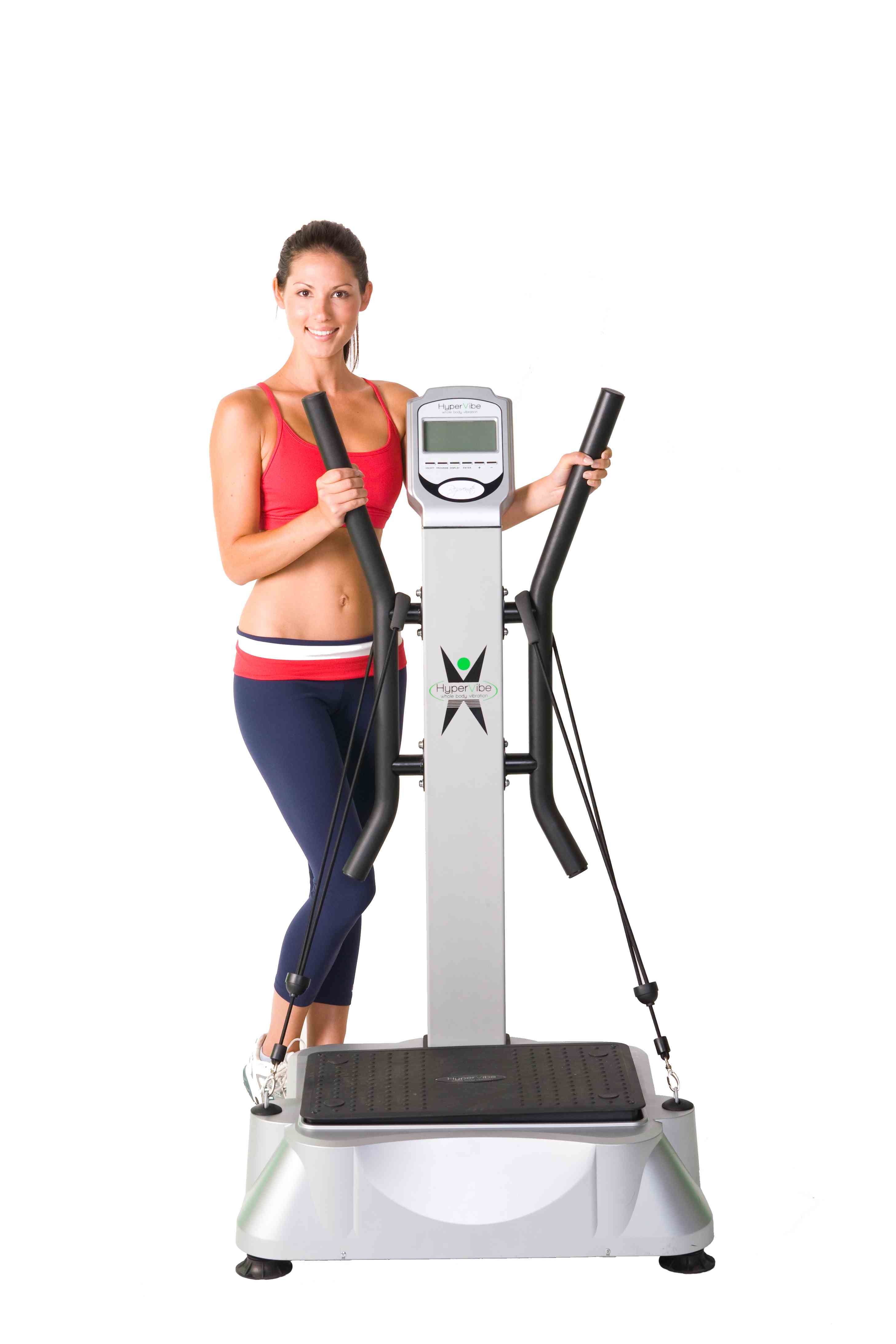
Newswise — Wounds may heal more quickly if exposed to low-intensity vibration, report researchers at the University of Illinois at Chicago.
The finding, in mice, may hold promise for the 18 million Americans who have type 2 diabetes, and especially the quarter of them who will eventually suffer from foot ulcers. Their wounds tend to heal slowly and can become chronic or worsen rapidly.
Timothy Koh, UIC professor of kinesiology and nutrition in the UIC College of Applied Health Sciences, was intrigued by studies at Stony Brook University in New York that used very low-intensity signals to accelerate bone regeneration.
“This technique is already in clinical trials to see if vibration can improve bone health and prevent osteoporosis,” Koh said.
Koh and his coworkers at UIC collaborated with Stefan Judex of Stony Brook to investigate whether the same technique might improve wound healing in diabetes. The new study, using an experimental mouse model of diabetes, is published online in the journal PLOS One.
The low-amplitude vibrations are barely perceptible to touch.
“It’s more like a buzz than an earthquake,” said Eileen Weinheimer-Haus, UIC postdoctoral fellow in kinesiology and nutrition, the first author of the study. The researchers found that wounds exposed to vibration five times a week for 30 minutes healed more quickly than wounds in mice of a control group.
Wounds exposed to vibration formed more granulation tissue, a type of tissue important early in the wound-healing process. Vibration helped tissue to form new blood vessels — a process called angiogenesis — and also led to increased expression of pro-healing growth factors and signaling molecules called chemokines, Weinheimer-Haus said.
“We know that chronic wounds in people with diabetes fail to form granulation tissue and have poor angiogenesis, and we believe these factors contribute to their wounds’ failure to heal,” said Koh.
He and his colleagues want to determine whether the changes they see in cell populations and gene expression at wound sites underlie the observed improvement in healing.
“The exciting thing about this intervention is how easily it could be translated to people,” Koh said.
“It’s a procedure that’s non-invasive, doesn’t require any drugs, and is already being tested in human trials to see if it’s protective of bone loss.” A clinical study, in collaboration with Dr. William Ennis, director of the Wound Healing Clinic at UIC, is planned, Koh said.
Ennis and Judex are co-authors on the PLOS One study, which was supported by National Institutes of Health grants R01GM092850 and T32DE018381.
- See more at: http://www.stonehearthnewsletters.com/vibration-may-help-heal-chronic-wounds/wound-care/#sthash.8fesSj1x.dpuf
Tuesday, April 8, 2014
Green tea said to boost brain: new research in psychopharmacology journal
Posted on April 7, 2014 by Stone Hearth News

Green tea is said to have many putative positive effects on health. Now, researchers at the University of Basel are reporting first evidence that green tea extract enhances the cognitive functions, in particular the working memory. The Swiss findings suggest promising clinical implications for the treatment of cognitive impairments in psychiatric disorders such as dementia. The academic journal Psychopharmacology has published their results.
In the past the main ingredients of green tea have been thoroughly studied in cancer research. Recently, scientists have also been inquiring into the beverage’s positive impact on the human brain. Different studies were able to link green tea to beneficial effects on the cognitive performance. However, the neural mechanisms underlying this cognitive enhancing effect of green tea remained unknown.
Better memory
In a new study, the researcher teams of Prof. Christoph Beglinger from the University Hospital of Basel and Prof. Stefan Borgwardt from the Psychiatric University Clinics found that green tea extract increases the brain’s effective connectivity, meaning the causal influence that one brain area exerts over another. This effect on connectivity also led to improvement in actual cognitive performance: Subjects tested significantly better for working memory tasks after the admission of green tea extract.
Green tea: our other posts
For the study healthy male volunteers received a soft drink containing several grams of green tea extract before they solved working memory tasks. The scientists then analyzed how this affected the brain activity of the men using magnetic resonance imaging (MRI). The MRI showed increased connectivity between the parietal and the frontal cortex of the brain. These neuronal findings correlated positively with improvement in task performance of the participants. «Our findings suggest that green tea might increase the short-term synaptic plasticity of the brain», says Borgwardt.
Clinical implications
The research results suggest promising clinical implications: Modeling effective connectivity among frontal and parietal brain regions during working memory processing might help to assess the efficacy of green tea for the treatment of cognitive impairments in neuropsychiatric disorders such as dementia.
Source - See more at: http://www.stonehearthnewsletters.com/green-tea-said-to-boost-brain-new-research-in-psychopharmacology-journal/green-tea-2/#sthash.IhzeT4bW.dpuf

Green tea is said to have many putative positive effects on health. Now, researchers at the University of Basel are reporting first evidence that green tea extract enhances the cognitive functions, in particular the working memory. The Swiss findings suggest promising clinical implications for the treatment of cognitive impairments in psychiatric disorders such as dementia. The academic journal Psychopharmacology has published their results.
In the past the main ingredients of green tea have been thoroughly studied in cancer research. Recently, scientists have also been inquiring into the beverage’s positive impact on the human brain. Different studies were able to link green tea to beneficial effects on the cognitive performance. However, the neural mechanisms underlying this cognitive enhancing effect of green tea remained unknown.
Better memory
In a new study, the researcher teams of Prof. Christoph Beglinger from the University Hospital of Basel and Prof. Stefan Borgwardt from the Psychiatric University Clinics found that green tea extract increases the brain’s effective connectivity, meaning the causal influence that one brain area exerts over another. This effect on connectivity also led to improvement in actual cognitive performance: Subjects tested significantly better for working memory tasks after the admission of green tea extract.
Green tea: our other posts
For the study healthy male volunteers received a soft drink containing several grams of green tea extract before they solved working memory tasks. The scientists then analyzed how this affected the brain activity of the men using magnetic resonance imaging (MRI). The MRI showed increased connectivity between the parietal and the frontal cortex of the brain. These neuronal findings correlated positively with improvement in task performance of the participants. «Our findings suggest that green tea might increase the short-term synaptic plasticity of the brain», says Borgwardt.
Clinical implications
The research results suggest promising clinical implications: Modeling effective connectivity among frontal and parietal brain regions during working memory processing might help to assess the efficacy of green tea for the treatment of cognitive impairments in neuropsychiatric disorders such as dementia.
Source - See more at: http://www.stonehearthnewsletters.com/green-tea-said-to-boost-brain-new-research-in-psychopharmacology-journal/green-tea-2/#sthash.IhzeT4bW.dpuf
Subscribe to:
Posts (Atom)